A Clearer View: Vascular Visualization Technologies for Easier IV Insertion
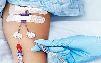
IV insertions can be one of the most challenging tasks in clinical care, especially when dealing with hard-to-stick patients. Fortunately, advancements in vascular visualization technology have transformed the way clinicians approach venipuncture—improving success rates, reducing patient discomfort, and saving time. Whether you’re a new nurse still mastering peripheral IV skills or a seasoned clinician looking to refresh your knowledge, understanding these tools is key.
Let’s break down the three primary types of vascular visualization tools used in today’s clinical settings: transilluminators, infrared devices, and ultrasound.
Transilluminators
Transilluminators use high-intensity LED light to pass through the skin and underlying tissue. Veins appear as dark lines against the illuminated background. This technique works best in pediatric patients or adults with fair or thin skin where veins are closer to the surface.

Pros:
- Simple and inexpensive
- Portable and easy to use
- No need for extensive training
Limitations:
- Less effective on adults with deep or darkly pigmented skin
- Not ideal for obese patients or those with significant edema
Infrared Technology (Near-Infrared Vein Finders)
Infrared vein finders use near-infrared light, which is absorbed by hemoglobin in the blood. The device captures this image and projects it back onto the skin in real time, revealing a map of subcutaneous veins.

Pros:
- Helpful for finding veins in difficult patients (e.g., obese, dark-skinned, dehydrated)
- Real-time projection helps guide insertion
- Non-invasive and user-friendly
Limitations:
- Less useful for deeper veins
- Cost can be a barrier for some facilities
- May require practice to interpret vein depth and trajectory
Ultrasound Guidance
Ultrasound-guided IV insertion uses high-frequency sound waves to provide a live image of veins beneath the skin. This method is especially helpful for deeper or less visible veins that can't be palpated or seen by other means.

Pros:
- Ideal for central lines and deep peripheral veins
- Improves success in patients with limited access
- Real-time visualization during cannulation
Limitations:
- Requires training and competency in vascular access with ultrasound
- Equipment is more expensive and less portable
- Not typically used for routine IV starts
Choosing the Right Tool
The decision to use a particular vascular visualization tool depends on the patient's condition, vein accessibility, and your facility's resources. These technologies are not replacements for good assessment and palpation skills—but they are powerful aids that can dramatically improve outcomes.
Want to Learn More?
For an in-depth review of these tools, when to use them, and how to incorporate them into your practice, check out our comprehensive Guide to Vascular Visualization. It’s packed with clinical tips, links to manufacturers guides, and practical considerations to take your IV insertion skills to the next level.